Catatonia is a complex and often perplexing condition that can leave both patients and clinicians in the dark. Characterized by motor immobilization, mutism, or agitation, it poses significant challenges for accurate diagnosis and effective management. One tool that has emerged as a beacon of hope in this field is electroencephalography (EEG). This non-invasive technique offers insights into brain activity, providing valuable data to help assess catatonic states.
Understanding the intricate relationship between EEG patterns and catatonia can unlock critical information for treatment pathways. As we delve deeper into how EEG aids in deciphering the enigmatic nature of catatonia, you’ll discover its essential role not just in diagnosis but also in crafting targeted therapeutic strategies. Join us on this journey to explore how advancements in EEG technology are reshaping our approach to one of psychiatry’s most challenging disorders.

Understanding EEG: Basic Principles and Relevance to Catatonia
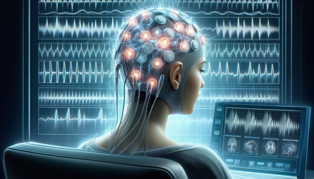
Electroencephalography (EEG) is a diagnostic tool that measures electrical activity in the brain through electrodes placed on the scalp. These electrodes detect voltage fluctuations resulting from neuron firing, creating a real-time map of brain wave patterns. This information is crucial for understanding various neurological and psychiatric conditions.
In the context of catatonia, EEG serves as a window into abnormal brain functioning. It helps clinicians identify specific neurophysiological changes associated with this condition. By analyzing these patterns, healthcare providers can gain insights into the severity and nature of catatonia.
The relevance of EEG in catatonia assessment cannot be overstated. Unlike other diagnostic methods, EEG is non-invasive and provides immediate feedback about brain activity. This makes it an invaluable resource during clinical evaluations when timely decisions are essential for patient care.
Moreover, advancements in technology have improved EEG’s sensitivity and specificity, enhancing its role in identifying subtle changes often overlooked by traditional assessments. Understanding these principles lays the groundwork for exploring how EEG contributes to diagnosing and managing catatonia effectively.
EEG Patterns in Catatonia: Characteristic Findings and Their Significance
Electroencephalography (EEG) reveals distinct patterns associated with catatonia, offering critical insights into this complex condition. One of the most notable findings is the prevalence of beta wave activity, which may indicate heightened arousal or anxiety levels in affected individuals. This increased beta activity often contrasts sharply with typical EEG patterns seen in other psychiatric disorders.
Another significant pattern is a decrease in alpha waves, suggesting impaired cortical functioning. Such alterations can help differentiate catatonia from other mental health issues and guide treatment strategies effectively. Observations like these underline how EEG serves as more than just a tool for monitoring brain activity; it plays an essential role in understanding underlying neural mechanisms.
Additionally, abnormal theta wave patterns have been reported during periods of immobilization common to catatonic states. These findings not only enhance diagnostic accuracy but also provide valuable information regarding potential neurophysiological triggers behind specific behaviors observed in patients.
The significance of these EEG characteristics extends beyond mere observation; they contribute to developing tailored interventions and improving patient outcomes through targeted therapies.
Quantitative EEG Analysis: Advanced Techniques for Catatonia Diagnosis
Quantitative EEG (qEEG) analysis represents a significant advancement in diagnosing catatonia. This method employs sophisticated algorithms to extract quantitative data from standard EEG recordings. By analyzing brain wave patterns, clinicians can identify abnormalities that may be indicative of catatonic states.
One of the primary benefits of qEEG is its ability to provide objective measurements. These metrics enable healthcare professionals to assess functional connectivity and identify specific oscillatory patterns associated with catatonia. Unlike traditional visual inspection, qEEG offers a more refined approach for understanding complex brain dynamics.
Moreover, certain frequency bands such as theta and delta have been shown to correlate with severity levels in catatonic patients. Tracking these changes over time can help gauge treatment response and adjust interventions accordingly.
As research continues in this area, integrating qEEG into clinical practice could enhance diagnostic accuracy. This promises improved patient outcomes by providing clearer insights into the underlying neurophysiology of catatonia.
EEG in Differential Diagnosis: Distinguishing Catatonia from Seizure Disorders
Electroencephalography (EEG) plays a vital role in distinguishing catatonia from seizure disorders. Both conditions can present with altered mental states, making accurate diagnosis essential for effective treatment. EEG offers a window into the brain’s electrical activity, revealing patterns that help differentiate these two complex disorders.
In catatonia, EEG findings may show generalized slowing or even normal activity. This contrasts sharply with seizure disorders, where spikes and sharp waves are typically observed on the EEG tracing. Recognizing these distinct patterns is crucial for clinicians aiming to initiate appropriate interventions.
Moreover, patients with catatonia might exhibit periods of unresponsiveness that can mimic seizures. Continuous monitoring through EEG helps clarify whether such episodes involve true epileptic activity or are part of the catatonic state.
By utilizing advanced EEG techniques alongside clinical assessments, healthcare providers can enhance diagnostic accuracy and avoid mislabeling patients. This differentiation not only guides treatment but also improves patient outcomes significantly.
Continuous EEG Monitoring: Benefits in Severe or Malignant Catatonia
Continuous EEG monitoring offers significant advantages for patients experiencing severe or malignant catatonia. This technique provides real-time insights into brain activity, allowing clinicians to observe any fluctuations or abnormalities that may occur over time. These persistent recordings can reveal patterns that might be missed during standard EEG assessments.
In cases of malignant catatonia, where symptoms can rapidly escalate, continuous monitoring is crucial. It enables timely interventions by identifying seizure-like activities or changes in brain wave patterns associated with significant distress. With this information at hand, healthcare providers can make informed decisions regarding treatment options.
Additionally, continuous EEG facilitates the assessment of medication responses in real time. Clinicians can closely evaluate how a patient’s brain responds to pharmacological treatments and adjust dosages accordingly for optimal management.
The ability to monitor neurological status continuously enhances overall patient safety and care quality. By keeping track of both clinical symptoms and electrical activity in the brain, healthcare teams are better equipped to manage complex cases effectively.
EEG Changes During Lorazepam Challenge Test in Catatonic Patients
The Lorazepam challenge test is a valuable tool in assessing catatonia, particularly when evaluating the response to benzodiazepines. Electroencephalography (EEG) plays a crucial role in observing these changes during the test. Characteristic EEG patterns can emerge as medication is administered.
In many cases, patients with catatonia demonstrate distinct shifts in brain wave activity post-Lorazepam administration. These alterations often manifest as increased theta and decreased alpha waves, indicating heightened cortical excitability. Such changes are essential for confirming a diagnosis of catatonia.
Furthermore, monitoring EEG responses allows clinicians to distinguish between different types of psychomotor agitation or immobility that may occur in various psychiatric disorders. The specificity of EEG findings helps tailor treatment approaches more effectively.
Understanding these EEG variations during the Lorazepam challenge enhances diagnostic accuracy and guides subsequent therapeutic decisions. This integration of neurophysiological insights marks an important advancement in managing complex cases of catatonia.
Limitations and Challenges of EEG in Catatonia Assessment
Electroencephalography (EEG) is a powerful tool, but it comes with limitations in assessing catatonia. One major challenge is the variability of EEG patterns among individuals. Not all patients exhibit clear or distinctive changes that can be easily interpreted, which complicates diagnosis.
Moreover, the presence of artifacts—such as muscle activity or eye movements—can interfere with readings. These extraneous factors may obscure significant brain wave patterns and lead to misinterpretations.
EEG’s time-sensitive nature also poses difficulties. Catatonic symptoms can fluctuate rapidly; thus, capturing relevant data during brief episodes becomes challenging. This limitation necessitates continuous monitoring for accurate assessments.
While EEG provides valuable insights into brain activity, it must be integrated with clinical evaluations for a comprehensive understanding of catatonia. Relying solely on EEG findings could result in incomplete assessments and hinder effective treatment strategies.
Integrating EEG Findings with Clinical Presentation: A Holistic Approach
Effectively assessing catatonia requires more than just EEG readings. Integrating these findings with clinical presentations provides a fuller picture of the patient’s condition. The nuances of behavior, mood, and physical health can heavily influence brain wave patterns observed in EEG.
Clinicians must pay attention to factors such as motor activity or lack thereof, responsiveness to external stimuli, and emotional states. These characteristics help in contextualizing the electrical activity recorded during an EEG session. For example, specific waveforms may appear distinctly different in patients experiencing acute catatonia versus those with underlying neurological disorders.
By combining symptoms and EEG data, healthcare providers can make informed decisions regarding treatment strategies. This holistic approach not only enhances diagnostic accuracy but also tailors interventions that address both physiological and psychological needs.
Collaboration among psychiatrists, neurologists, and other specialists is essential for integrating these diverse elements effectively. Such teamwork fosters comprehensive care plans that improve outcomes for patients facing complex challenges associated with catatonia.
Emerging EEG Technologies: Future Directions in Catatonia Diagnosis
Recent advancements in EEG technology are paving new paths for catatonia diagnosis. High-density EEG systems, which use numerous electrodes, provide a more detailed view of brain activity. This enhanced resolution may reveal subtle changes that traditional EEG methods might miss.
Machine learning algorithms are also making waves in the analysis of EEG data. These intelligent systems can identify patterns and anomalies associated with catatonia, potentially speeding up diagnosis and improving accuracy. As these technologies evolve, their integration into clinical settings could transform how specialists assess patients.
Wearable EEG devices represent another innovation on the horizon. These portable tools allow for continuous monitoring outside hospital environments. Such flexibility could lead to earlier detection of catatonic symptoms in real-world settings.
Moreover, combining EEG with neuroimaging techniques such as fMRI offers a comprehensive approach to understanding brain function during episodes of catatonia. This multimodal strategy promises richer insights into the underlying mechanisms at play in this complex condition.
EEG-Guided Treatment Strategies in Catatonia Management
EEG-guided treatment strategies are emerging as a pivotal component in managing catatonia. By leveraging the insights gained from electroencephalography, clinicians can tailor interventions more effectively to address the specific needs of each patient.
The data obtained through EEG allows for real-time monitoring of brain activity during treatment. This is particularly valuable when administering pharmacological agents like benzodiazepines or anesthetics, which may significantly alter brain wave patterns. Understanding these changes helps healthcare providers assess the effectiveness of treatments and make timely adjustments if necessary.
Additionally, EEG findings can inform non-pharmacological approaches such as neuromodulation techniques. These methods hold promise in cases where traditional therapies fall short. The integration of EEG into treatment planning enhances not only diagnostic accuracy but also therapeutic outcomes.
As research advances, we will likely see an increased emphasis on personalized medicine in catatonia management. Harnessing the crucial role of electroencephalography (EEG) in catatonia assessment opens new avenues for improving care and ultimately enhancing patients’ quality of life through informed and targeted interventions.


